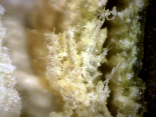
This neighborhood White Oak didn't look healthy at all. So I clipped a branch from
it and decided to check it out microscopically. The branch sample was obtained in late
September. The results are shown below.
1
This tangled mass of canker material is pushing out the outer bark, which is also riddled with canker.
The canker material ranges in color from tan to white. (400x)
 ↓
↓
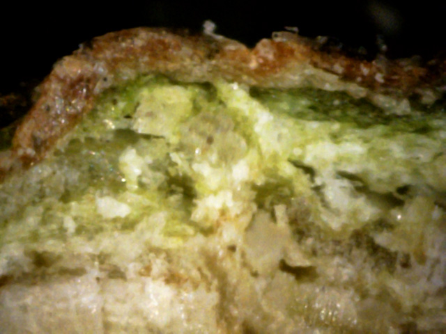
2
Note the "D" shaped void (red arrow) - probably caused by the pressure of the surrounding canker growth.
The canker outside the void is thicker than the inside, which probably pushed it out further
than the inside could push in. Plus, the green sapwood appears riddled with white canker particles.
(400x)
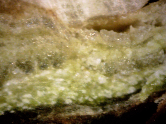
3
The mangled sapwood tissue in this view contains canker material plus lots of hyphae.
Even bark surface canker can be seen. (400x)
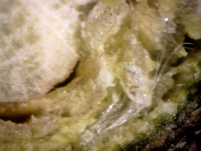
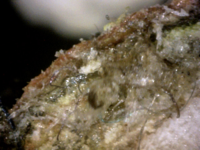
4
There isn't much left of the green sapwood.
Two thick bands of canker growth have expanded to the point where they both left
"D" shaped voids in the wood.
And one bundle of crystal-clear hyphae reaches up through a void and spreads out. (400x)
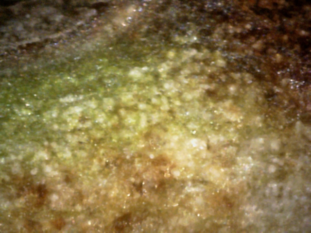
6
When doing a twig cross-section, I had always wondered if the small white circular blobs
were really spherical or the ends of a circular tube.
This view was taken with a razor cut of about 10 degrees from the twig axis.
If the blobs were really tubes, we would see many long ovals.
Instead we still see circles, meaning that the small white blobs are really pretty spherical in shape. (400x)
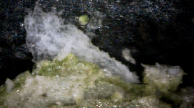
7
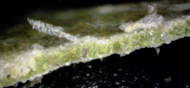
A good leaf cross-section view that shows two different kinds of canker material.
The large blob at the top is mostly filmy and transparent, while the blob at the bottom
is more gray in color.
Canker that is separated from leaf material tends to be snow white or transparent. (400x)
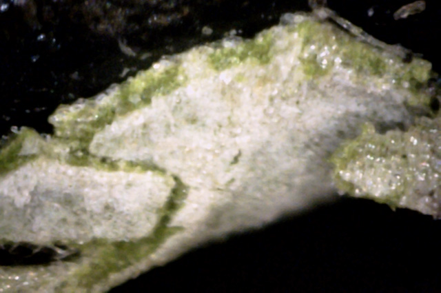 ↓
←
↓
←
8
A good view of a leaf cross-section showing both the inner chlorophyl (blue arrow) and
the leaf bottom that is covered with this white canker material.
The surface rip in the leaf bottom (red arrow) shows how dense this canker material is, and how completely it covers
the leaf bottom, probably choking off the leaf. (400x)
 ↓
↓
←
↓
↓
←
9
This leaf cross-section view also shows us some of the top of the leaf.
The two blobs of canker on the top resemble bubble-wrap (blue arrows).
Click the picture to examine the left one in detail - a hypha came up through
the leaf and its tendrils engulfed some leaf cells, digesting them.
A chunk of gray canker (red arrow) material sits on the leaf at the left of the picture. (400x)
 ↓
↓
10
Here again the leaf bottom is covered with a dense coating of white material,
and the top is also covered. But the shapes and transparency of the canker differs.
A leaf vein (red arrow) is on the left. (400x)
 ↓
↓
11
The bottom of this leaf is totally covered with what looks like white canker.
A vein is on the left. The leaf top supports numerous clear canker material (the
red arrows points to one blob). (400x)
 →
→
12
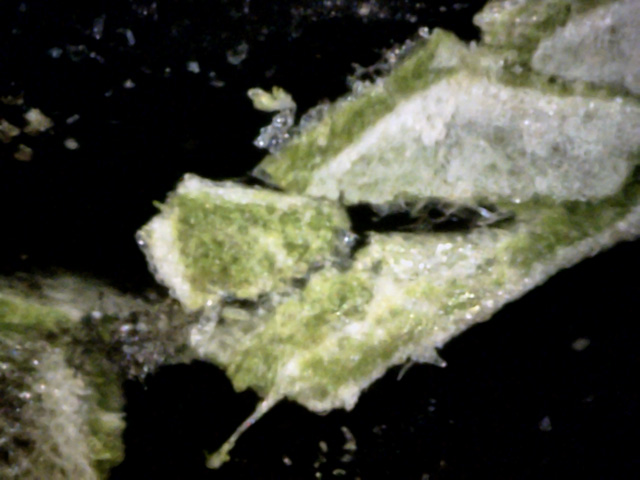
I'd always wanted to cut a leaf through its inside so I could see between the sides,
as in removing the slices of bread making up a sandwich.
By luck, the razor I used to make this cross-section tore open the leaf for me here!
A long canker hypha can be seen (red arrow) running along the leaf, and then continuing on out of the leaf.
Apparently it is more cohensive than the leaf cells. The bottom of this view is the leaf bottom (400x)
As the pictures show, there is no question that this white oak is severely infected with white canker.
The extent of this infection makes one realize how tough trees are. And, this severe infection did illustrate
several additional details about the white canker disease.
